Iso-acoustic focusing
Analyzing cells' acoustic properties
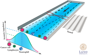
In iso-acoustic focusing (IAF), individual cells are analyzed based on the previously uncharted parameter of effective acoustic impedance [Augustsson et al., Nature Communications, 2016]. This equilibrium method can be viewed as a microfluidic analog to density gradient centrifugation or iso-electric focusing. Cells flowing through a microchannel migrate sideways, influenced by an acoustic field, into flow streams of ever increasing acoustic impedance. Finally, the individual cells reach their iso-acoustic point (IAP), at which the acoustic contrast between the cell and the surrounding liquid becomes zero, and the sideways displacement ceases. Cell-specific differences in effective acoustic impedance translate to a spatial dispersion of the cell population transverse to the flow, enabling continuous label-free analysis of individual cells.
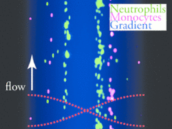
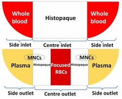
Separating cells based on acoustic properties
By this approach, we recently presented the first one-step acoustic separation of mononuclear cells (MNCs) from undiluted human whole blood [Alsved et al. Microtas, Basel, 2019 (PDF)].